|
产品名称 |
LCLC-103H人大细胞肺癌 |
|
货号 |
ZQ1153 |
|
产品介绍 |
LCLC-103H是一种人类大细胞肺癌(Large Cell Lung Cancer, LCLC)细胞系。大细胞肺癌是一种非小细胞肺癌(Non-Small Cell Lung Cancer, NSCLC)的亚型,建立于一个61岁的高加索大细胞肺癌患者的胸腔积液,是一种未分化的肺癌,并有巨大的细胞,他接受了化疗和放疗。该细胞具有快速生长和侵袭性的特点。这种类型的肺癌细胞在显微镜下观察时,通常呈现出较大的不规则形状,且细胞核较大。文献中描述这是阴性的,表现出显著的基质形成,并过度压迫原癌基因肌瘤。该细胞系特别值得注意的是其神经内分泌标记的部分表达,这些标记通常与小细胞肺癌(SCLC)和某些神经内分泌肿瘤有关。特别地,由单克隆抗体RNL-1检测的抗原在LCLC-103H细胞中显示出病灶表面表达,类似于在一些神经内分泌癌中观察到的。然而,所有细胞中的表达并不一致,表明细胞群体中的异质性。
LCLC-103H在文献中被描述为PAS(高碘酸-希夫)阴性,将其与其他肺癌亚型相区别。它还表现出显著的基质形成,这是其组织病理学特征的显著特征。此外,已知该细胞系过度表达在细胞增殖和肿瘤发生中起关键作用的原癌基因MYC。免疫细胞化学研究表明,LCLC-103H不显示小细胞肺癌中所见的全部神经内分泌分化,因为它缺乏与其他神经内分泌标记物的反应性,例如由抗体RNL-2和RNL-3鉴定的那些。这一区别对于区分LCLC和小细胞肺癌至关重要,小细胞肺癌更具侵袭性,通常对某些化疗药物表现出更高的敏感性。LCLC-103H的独特表达谱使其成为研究大细胞肺癌的分子和免疫学特征及其与神经内分泌特征重叠的有价值的模型。 |
|
种属 |
人 |
|
性别/年龄 |
男/61岁 |
|
组织 |
肺 |
|
疾病 |
大细胞肺癌 |
|
细胞类型 |
肿瘤细胞 |
|
形态学 |
上皮 |
|
生长方式 |
贴壁 |
|
倍增时间 |
大约26~40 小时 |
|
培养基和添加剂 |
RPMI-1640(品牌:中乔新舟 货号:ZQ-200)+10%胎牛血清(中乔新舟 货号:ZQ500-A)+1%P/S(中乔新舟 货号:CSP006) |
|
推荐完全培养基货号 |
|
|
生物安全等级 |
BSL-1 |
|
培养条件 |
95%空气,5%二氧化碳;37℃ |
|
STR位点信息 |
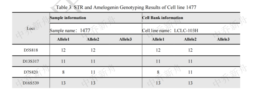
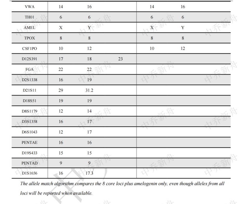
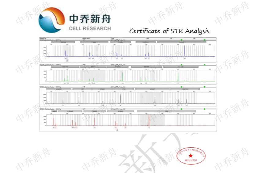
Amelogenin :X (CLS=300169) |
|
抗原表达/受体表达 |
*** |
|
基因表达 |
*** |
|
保藏机构 |
CLS; 300169 DSMZ; ACC-384 |
|
供应限制 |
仅供科研使用 |
|
货号 |
ZQ1153 |
|
发货规格 |
活细胞:T25培养瓶*1瓶或者1ml 冻存管*2支(细胞量约为1x10^6 Cells/Vial )二选一 |
|
发货形式 |
活细胞:常温运输;冻存管:干冰运输 |
|
储存温度 |
活细胞:培养箱;冻存管:液氮罐 |
|
产地 |
中国 |
|
供应限制 |
仅供科研使用 |
PubMed=2473086; DOI=10.1242/jcs.91.1.91
Broers J.L.V., Rot M.K., Oostendorp T., Bepler G., de Leij L.F.M.H., Carney D.N., Vooijs G.P., Ramaekers F.C.S.
Spontaneous changes in intermediate filament protein expression patterns in lung cancer cell lines.
J. Cell Sci. 91:91-108(1988)
PubMed=2840315; DOI=10.1111/j.1432-0436.1988.tb00806.x
Bepler G., Koehler A., Kiefer P., Havemann K., Beisenherz K., Jaques G., Gropp C., Haeder M.
Characterization of the state of differentiation of six newly established human non-small-cell lung cancer cell lines.
Differentiation 37:158-171(1988)
PubMed=1845952; DOI=10.1002/1097-0142(19910201)67:3<619::AID-CNCR2820670317>3.0.CO;2-Y
Broers J.L.V., Mijnheere E.P., Rot M.K., Schaart G., Sijlmans A., Boerman O.C., Ramaekers F.C.S.
Novel antigens characteristic of neuroendocrine malignancies.
Cancer 67:619-633(1991)
PubMed=20215515; DOI=10.1158/0008-5472.CAN-09-3458
Rothenberg S.M., Mohapatra G., Rivera M.N., Winokur D., Greninger P., Nitta M., Sadow P.M., Sooriyakumar G., Brannigan B.W., Ulman M.J., Perera R.M., Wang R., Tam A., Ma X.-J., Erlander M., Sgroi D.C., Rocco J.W., Lingen M.W., Cohen E.E.W., Louis D.N., Settleman J., Haber D.A.
A genome-wide screen for microdeletions reveals disruption of polarity complex genes in diverse human cancers.
Cancer Res. 70:2158-2164(2010)
PubMed=22460905; DOI=10.1038/nature11003
Barretina J.G., Caponigro G., Stransky N., Venkatesan K., Margolin A.A., Kim S., Wilson C.J., Lehar J., Kryukov G.V., Sonkin D., Reddy A., Liu M., Murray L., Berger M.F., Monahan J.E., Morais P., Meltzer J., Korejwa A., Jane-Valbuena J., Mapa F.A., Thibault J., Bric-Furlong E., Raman P., Shipway A., Engels I.H., Cheng J., Yu G.-Y.K., Yu J.-J., Aspesi P. Jr., de Silva M., Jagtap K., Jones M.D., Wang L., Hatton C., Palescandolo E., Gupta S., Mahan S., Sougnez C., Onofrio R.C., Liefeld T., MacConaill L.E., Winckler W., Reich M., Li N.-X., Mesirov J.P., Gabriel S.B., Getz G., Ardlie K., Chan V., Myer V.E., Weber B.L., Porter J., Warmuth M., Finan P., Harris J.L., Meyerson M.L., Golub T.R., Morrissey M.P., Sellers W.R., Schlegel R., Garraway L.A.
The Cancer Cell Line Encyclopedia enables predictive modelling of anticancer drug sensitivity.
Nature 483:603-607(2012)
PubMed=25485619; DOI=10.1038/nbt.3080
Klijn C., Durinck S., Stawiski E.W., Haverty P.M., Jiang Z.-S., Liu H.-B., Degenhardt J., Mayba O., Gnad F., Liu J.-F., Pau G., Reeder J., Cao Y., Mukhyala K., Selvaraj S.K., Yu M.-M., Zynda G.J., Brauer M.J., Wu T.D., Gentleman R.C., Manning G., Yauch R.L., Bourgon R., Stokoe D., Modrusan Z., Neve R.M., de Sauvage F.J., Settleman J., Seshagiri S., Zhang Z.-M.
A comprehensive transcriptional portrait of human cancer cell lines.
Nat. Biotechnol. 33:306-312(2015)
PubMed=25877200; DOI=10.1038/nature14397
Yu M., Selvaraj S.K., Liang-Chu M.M.Y., Aghajani S., Busse M., Yuan J., Lee G., Peale F.V., Klijn C., Bourgon R., Kaminker J.S., Neve R.M.
A resource for cell line authentication, annotation and quality control.
Nature 520:307-311(2015)
PubMed=26589293; DOI=10.1186/s13073-015-0240-5
Scholtalbers J., Boegel S., Bukur T., Byl M., Goerges S., Sorn P., Loewer M., Sahin U., Castle J.C.
TCLP: an online cancer cell line catalogue integrating HLA type, predicted neo-epitopes, virus and gene expression.
Genome Med. 7:118.1-118.7(2015)
PubMed=27397505; DOI=10.1016/j.cell.2016.06.017
Iorio F., Knijnenburg T.A., Vis D.J., Bignell G.R., Menden M.P., Schubert M., Aben N., Goncalves E., Barthorpe S., Lightfoot H., Cokelaer T., Greninger P., van Dyk E., Chang H., de Silva H., Heyn H., Deng X.-M., Egan R.K., Liu Q.-S., Mironenko T., Mitropoulos X., Richardson L., Wang J.-H., Zhang T.-H., Moran S., Sayols S., Soleimani M., Tamborero D., Lopez-Bigas N., Ross-Macdonald P., Esteller M., Gray N.S., Haber D.A., Stratton M.R., Benes C.H., Wessels L.F.A., Saez-Rodriguez J., McDermott U., Garnett M.J.
A landscape of pharmacogenomic interactions in cancer.
Cell 166:740-754(2016)
PubMed=30894373; DOI=10.1158/0008-5472.CAN-18-2747
Dutil J., Chen Z.-H., Monteiro A.N.A., Teer J.K., Eschrich S.A.
An interactive resource to probe genetic diversity and estimated ancestry in cancer cell lines.
Cancer Res. 79:1263-1273(2019)
PubMed=31068700; DOI=10.1038/s41586-019-1186-3
Ghandi M., Huang F.W., Jane-Valbuena J., Kryukov G.V., Lo C.C., McDonald E.R. III, Barretina J.G., Gelfand E.T., Bielski C.M., Li H.-X., Hu K., Andreev-Drakhlin A.Y., Kim J., Hess J.M., Haas B.J., Aguet F., Weir B.A., Rothberg M.V., Paolella B.R., Lawrence M.S., Akbani R., Lu Y.-L., Tiv H.L., Gokhale P.C., de Weck A., Mansour A.A., Oh C., Shih J., Hadi K., Rosen Y., Bistline J., Venkatesan K., Reddy A., Sonkin D., Liu M., Lehar J., Korn J.M., Porter D.A., Jones M.D., Golji J., Caponigro G., Taylor J.E., Dunning C.M., Creech A.L., Warren A.C., McFarland J.M., Zamanighomi M., Kauffmann A., Stransky N., Imielinski M., Maruvka Y.E., Cherniack A.D., Tsherniak A., Vazquez F., Jaffe J.D., Lane A.A., Weinstock D.M., Johannessen C.M., Morrissey M.P., Stegmeier F., Schlegel R., Hahn W.C., Getz G., Mills G.B., Boehm J.S., Golub T.R., Garraway L.A., Sellers W.R.
Next-generation characterization of the Cancer Cell Line Encyclopedia.
Nature 569:503-508(2019)
PubMed=31978347; DOI=10.1016/j.cell.2019.12.023
Nusinow D.P., Szpyt J., Ghandi M., Rose C.M., McDonald E.R. III, Kalocsay M., Jane-Valbuena J., Gelfand E.T., Schweppe D.K., Jedrychowski M.P., Golji J., Porter D.A., Rejtar T., Wang Y.K., Kryukov G.V., Stegmeier F., Erickson B.K., Garraway L.A., Sellers W.R., Gygi S.P.
Quantitative proteomics of the Cancer Cell Line Encyclopedia.
Cell 180:387-402.e16(2020)
PubMed=35839778; DOI=10.1016/j.ccell.2022.06.010
Goncalves E., Poulos R.C., Cai Z.-X., Barthorpe S., Manda S.S., Lucas N., Beck A., Bucio-Noble D., Dausmann M., Hall C., Hecker M., Koh J., Lightfoot H., Mahboob S., Mali I., Morris J., Richardson L., Seneviratne A.J., Shepherd R., Sykes E., Thomas F., Valentini S., Williams S.G., Wu Y.-X., Xavier D., MacKenzie K.L., Hains P.G., Tully B., Robinson P.J., Zhong Q., Garnett M.J., Reddel R.R.
Pan-cancer proteomic map of 949 human cell lines.
Cancer Cell 40:835-849.e8(2022)


 上海中乔新舟生物科技有限公司